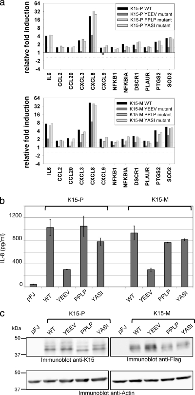
FIG. 4.
Contribution of conserved K15 SH2 and SH3 binding motifs to inflammatory signaling. (a) HeLa cells were transfected with expression vectors for the K15-P or K15-M wild-type (WT) protein or its mutant YEEV, PPLP, or YASI versions. Extracted RNA was subjected to low-density microarray analysis using a Human Inflammation Array (Ocimum Biosolutions). Ratios of relative gene expression were calculated by dividing signal intensity values originating from K15-P (top), K15-M (bottom), or mutant protein samples by values obtained for the empty vector control. The graphs show ratio values for genes that were induced at least 1.5-fold by both the K15-P WT and the K15-M WT proteins. (b) HeLa cells were transfected with expression vectors for the K15-P or K15-M WT or its mutant YEEV, PPLP, or YASI versions, as described for low-density microarrays, and IL-8 levels (pg/ml) in cleared supernatants were measured by ELISA. (c) Cleared lysates from transfected HeLa cells were analyzed with an anti-K15 antibody for equal K15-P expression, an anti-Flag antibody for equal K15-M expression, and an anti-actin antibody as a control.

