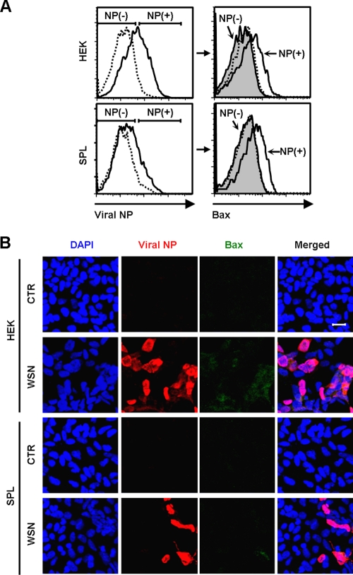
FIG. 3.
Coexpression of Bax and viral NP in influenza virus-infected HEK or SPL cells. (A) HEK cells or SPL cells were either left uninfected (dotted lines) or infected (solid lines) with WSN virus at an MOI of 1. (Left) At 2 dpi, the expression of viral NP was assessed by flow cytometry. Virus-treated cells (solid lines) were separated into NP-expressing (NP+) and nonexpressing (NP−) cells. (Right) The Bax expression of the NP+ cells (solid lines) and the NP− cells (shaded areas) was compared with that of uninfected cells (dotted lines). (B) Cells were either left uninfected (CTR) or infected with WSN at an MOI of 1. They were fixed, permeabilized, and stained with DAPI, to detect nuclei, and with antibodies against viral NP and Bax at 2 dpi. Representative confocal images are shown (original magnification, ×200). Bar, 20 μm.

