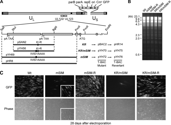
FIG. 3.
Generation of the IE2 SIM mutant and its revertant HCMV (Towne) BAC clones and their infectivities in transfected HF. (A) Scheme for the generation of the IE2 SIM mutant (mSIM) Towne (T)-BACs and their revertant T-BAC clones. A pGS284 (38) derivative (pYH69), which harbors a 4.1-kb DNA fragment containing the mSIM allele of IE2, was used for homologous recombination with the parental T-BAC clone. This recombination event resulted in the IE2(mSIM) T-BAC clone (pYH72). Similarly, the IE2(KR/mSIM) T-BAC clone (pYH70) was produced using a GS284 derivative (pYH56) containing a 2.4-kb IE2 KR/mSIM allele. A pGS284 derivative (pHR8) containing a 4.1-kb wild-type DNA fragment was used to make their revertant T-BAC clones (pYH76 and pYH75). The IE2 KR mutant T-BAC (pBAC2) and its revertant (pHR14) have been described previously (32). (B) Restriction fragment DNA patterns obtained following EcoRI/BamHI digestion of the wild-type, mutant, and revertant T-BAC DNAs were analyzed by agarose gel electrophoresis. λ-HindIII was used as the molecular weight standard. (C) Infectivities of the transfected T-BAC DNAs. HF were transfected with T-BAC clones (Wt, mSIM [pYH72], mSIM-R [pYH76], KR/mSIM [pYH70], and KR/mSIM-R [pYH75]) via electroporation (see Materials and Methods). The cells were monitored for the spread of GFP signals. GFP images (top) and their phase-contrast images (bottom) were photographed 4 weeks after electroporation. Representative images from at least four independent experiments are shown. The inserts in the images of mSIM virus infection include some GFP-positive cells. Note that the spread of these GFP signals in cells transfected with the mSIM and KR/mSIM T-BAC DNAs was usually incomplete compared to the spread in cells transfected with the wild-type or revertant T-BAC DNAs.

