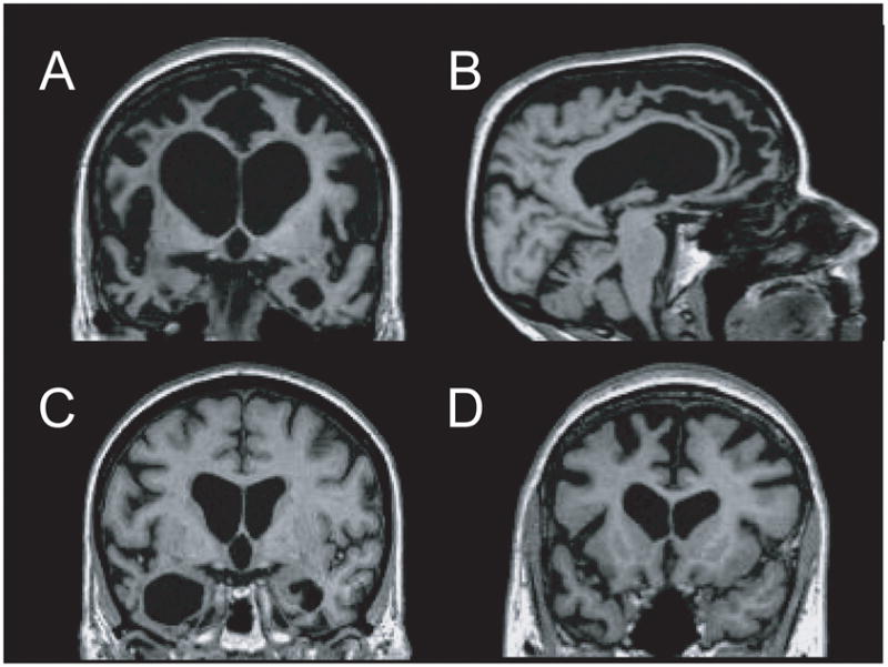
Fig. 2.

MRI findings in frontotemporal lobar degeneration (FTLD). T1-weighted images from representative patients with behavioural-variant frontotemporal dementia (bvFTD) [a and b], semantic dementia (SD) [c] and progressive nonfluent aphasia (PNFA) [d] are displayed in neurologic orientation. (a and b) bvFTD patient shows marked atrophy throughout the medial and lateral frontal cortex and the temporal poles, with striking relative preservation of the posterior brain regions on a sagittal view. (c) Patient with SD shows asymmetric degeneration of the temporal poles (left greater than right). (d) PNFA patient shows atrophy in the left inferolateral and dorsomedial frontal cortex and anterior insula.
