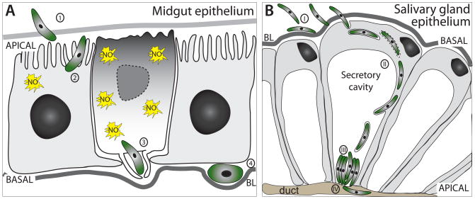
Fig. 1.
Comparison of malaria parasite entry into midgut and salivary gland epithelia. A) Ookinete invasion of midgut epithelia. The ookinete crosses the peritrophic matrix (1) and actively enters the epithelial cell 24–28 h after a bloodmeal, often sequentially invading several midgut cells (2). The ookinete egresses the epithelial cell at the basal site, which is accompanied by lamellipodia formation by the invaded cell as well as its neighbors (3). In the extracellular space of the basal labyrinth the ookinete starts to round up beneath the basal lamina (BL) and transforms into the oocyst (4). The invaded midgut cells undergo many severe physiological changes, among those nitric oxide (NO) production. Ultimately, the cells undergo apoptosis and are expelled from the epithelium. B) Salivary gland infection by sporozoites can be divided into several stages: (I) Sporozoite attachment, likely due to receptor-ligand interactions; (II) sporozoite invasion of epithelial cells with the formation of a transient vacuole; and (III) maturation phase, where sporozoites rest in the extracellular space of the secretory cavity, while (IV) few enter the salivary duct.

