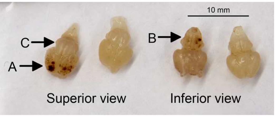
Figure 4. Hemorrhage in fetal brains.
Photographs of brains from SVCT2(−/−) (left-hand example of each pair) and SVCT2(+/+) fetuses from above (superior) and below (inferior), showing severe hemorrhage in cortex (A) and brain stem (B), and lack of hemorrhage in the cerebellum (C) in SVCT2(−/−) mice.

