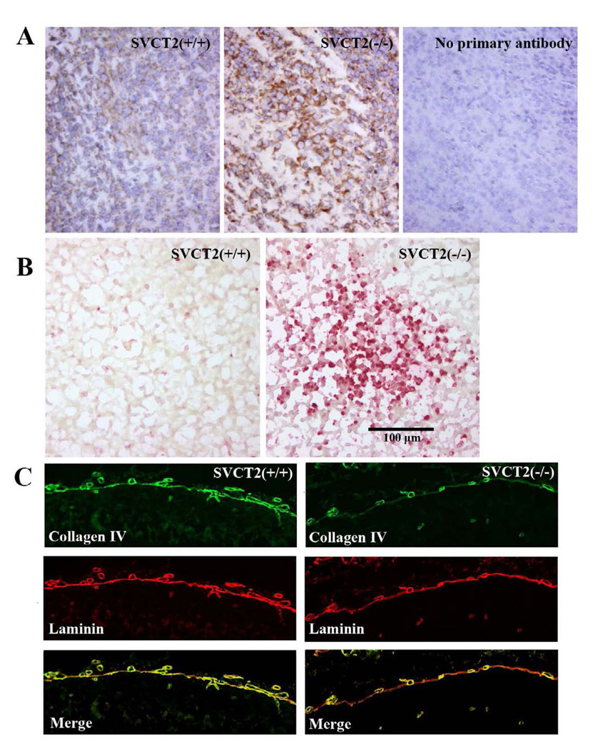
Figure 6. Isoketal staining, apoptosis and type IV collagen in late-stage SVCT2(−/−) and SVCT2(+/+) fetuses.
(A) Isoketal immunostaining was conducted with D11 ScFv antibody in mouse cortex and counterstained with hematoxylin and eosin. Greater isoketal staining was observed in SVCT2(−/−) embryos (center panel), compared to SVCT2(+/+) fetuses (left panel). The right panel shows no primary antibody (20x magnification). (B) Localized accumulations of TUNEL-positive cells were observed in cortex of SVCT2(−/−) fetuses compared to age-matched wild-type fetuses (20x magnification). (C) Double-immunofluorescent detection of collagen IV (green) and laminin (red) was conducted in brain of SVCT2(+/+) and SVCT2(−/−) fetuses. Note collagen IV is co-localized with laminin in the basement membranes of wild type (left panel) but not SVCT2(−/−) vascular vessels (right panel) (20x magnification).

