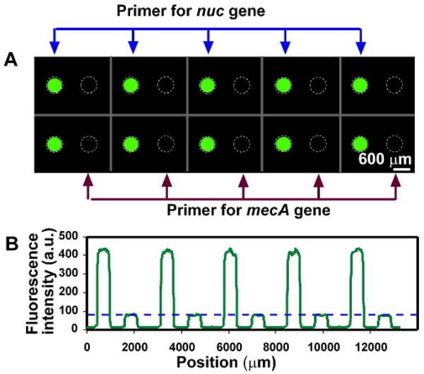
Figure 4.

No cross-contamination among adjacent wells was seen after PCR on the SlipChip. Primer pairs for the nuc gene in S. aureus and the mecA gene in Methicillin-resistant S. aureus (MRSA) were preloaded on the chip alternatively in the same row. PCR master mixture, containing 100 pg/μL of S. aureus (MSSA) genomic DNA, was injected into the SlipChip. A) Fluorescent microphotograph shows that only wells preloaded primers for the nuc gene (left in each pair of wells) showed fluorescent signal corresponding to DNA amplification. B) A line scan across the middle of the top row in (A) shows that fluorescence intensity of wells loaded with nuc primers were more than 5 fold higher than that of the wells loaded with mecA primers (p < 0.0001, n = 10) The blue dashed line in (B) represents the fluorescence intensity before thermal cycling in the wells.
