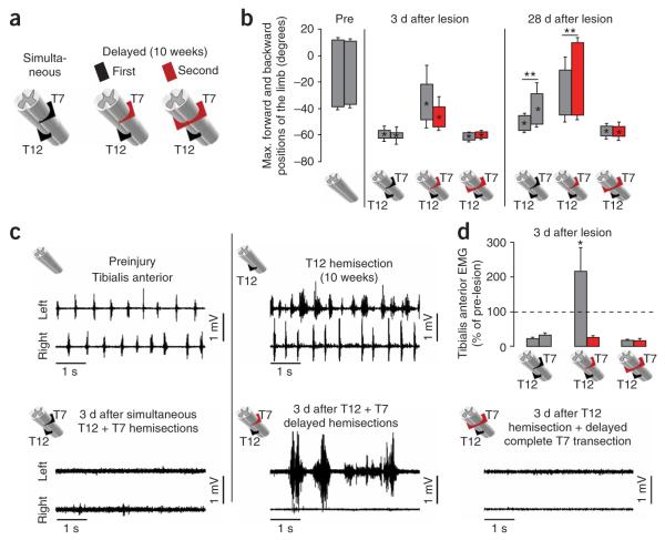
Figure 3.
Recovery of supraspinal control of stepping after delayed but not simultaneous T12 (left) and T7 (right) lateral hemisections. (a) Schematic of different combinations of simultaneous or delayed (10 weeks) SCIs. (b) Bar graphs showing the average maximum backward and forward angular positions of the limb axis after different SCIs. (c) Representative EMG recordings from the left and right tibialis anterior during stepping before and 3 d after different SCIs. (d) Bar graphs showing the average EMG burst amplitude for left and right tibialis anterior muscles (n = 4 for simultaneous or delayed hemisections; n = 3 for complete delayed T7 transection). Values are normalized to prelesion EMG activity. Values represent means ± s.e.m. *P < 0.05 and **P < 0.01, statistically significant differencs between pre- and postlesion values and between ipsilateral and contralateral hindlimbs, respectively.

