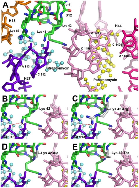
Figure 3. Three-dimensional crystal structure of T. thermophilus ribosomal decoding A-site with bound paromomycin and streptomycin.
(A) General view of the A-site. Amino-acid residues of S12 (green) are shown labelled according to atoms: carbon – green, nitrogen – blue, oxygen – red. 16S rRNA helices are indicated as follows: H44 strand I (pink), H44 strand II (magenta), H27 (violet), H18 (orange). Streptomycin (light blue) and paromomycin (yellow). (B) Close up of wild type K42. Hydrogen bonds to streptomycin (black dotted lines) and salt bridge to A913 (red dotted line) are shown. (C) Close up of mutant K42R. Salt bridge (red dotted line) is shown. (D) Close up of mutant K42A. (E) Close up of mutant K42T. (Protein Data Bank, 1FJG.pdb).

