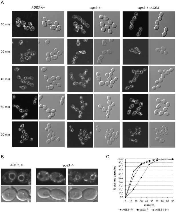
Figure 3. Cells lacking AGE3 show a clear delay in endocytosis.
(A) Cells of the strains SN87 (AGE3+/+), UZ45 (age3Δ/Δ) and UZ55 (age3Δ/Δ::AGE3) were grown in YPD to exponential phase. After staining the cytoplasmic membrane with the lipophilic fluorescent dye FM4-64 at 0°C for 40 minutes, the excess dye was removed by washing. Then the cells were released for growth in YPD at 30°C to allow endocytosis to occur. Samples were taken at the indicated time points. Staining of endocytic vesicles and the vacuole was visualized by confocal fluorescence microscopy. DIC images are shown for comparison. (B) One and two cell pairs of the wild-type and the age3Δ/Δ mutant strains, respectively, which were harvested 40 minutes after release of endocytosis are shown in higher magnification. The mutant cells show stronger background staining of the cytoplasm and more bright spots compared to the wild-type cells. The vauolar membranes of the left mutant cells still have not taken up FM4-64. (C) Quantification of FM4-64 uptake into vacuolar membranes in cells of the strains mentioned above. Images of a similar experiment as described under (A) were analysed and the percentage of cells (about 100–150 in total for each strain and time point) with clearly stained vacuolar membranes determined. A similar experiment performed on another day with slightly different time points showed a very similar FM4-64 uptake kinetics.

