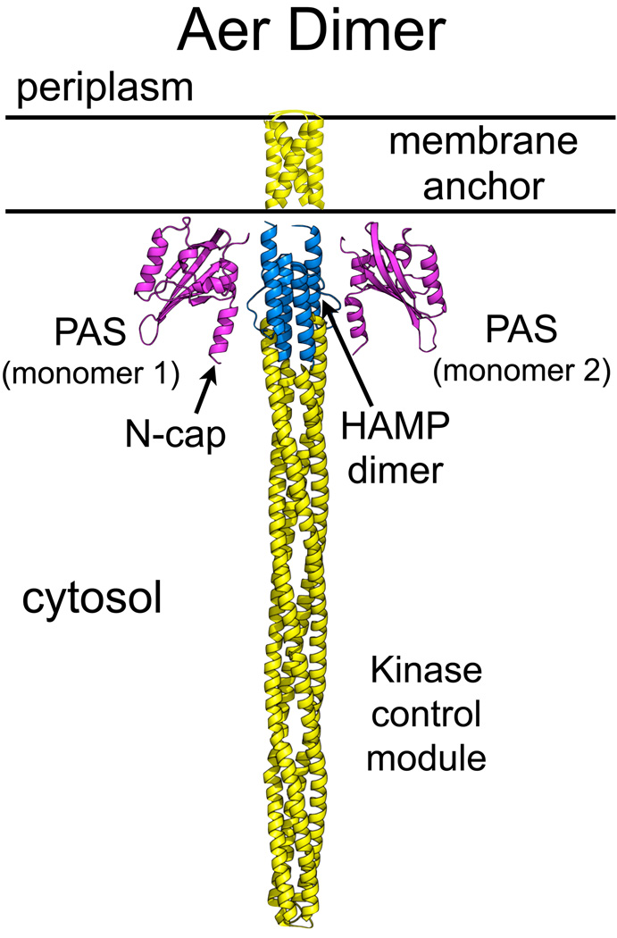
Fig. 1.
Model of the domain organization of an Aer dimer. Structures were modeled in silico with Swiss-Model (Kiefer et al., 2009) based on PAS-A from AvNifL [2GJ3 (Key et al., 2007); Aer-PAS: cyan], Af1503 HAMP [2ASW (Hulko et al., 2006); Aer-HAMP: blue] and the cytoplasmic domain from a Thermatoga maritima receptor [2CH7 (Park et al., 2006); Aer-kinase control module: yellow]. The membrane anchor is based on disulfide crosslinking studies [yellow, (Amin et al., 2007, Amin et al., 2006)]. The PAS domains are linked to the membrane anchor via a forty-five amino acid F1 tether (not drawn); the HAMP domain begins immediately following the membrane anchor.

