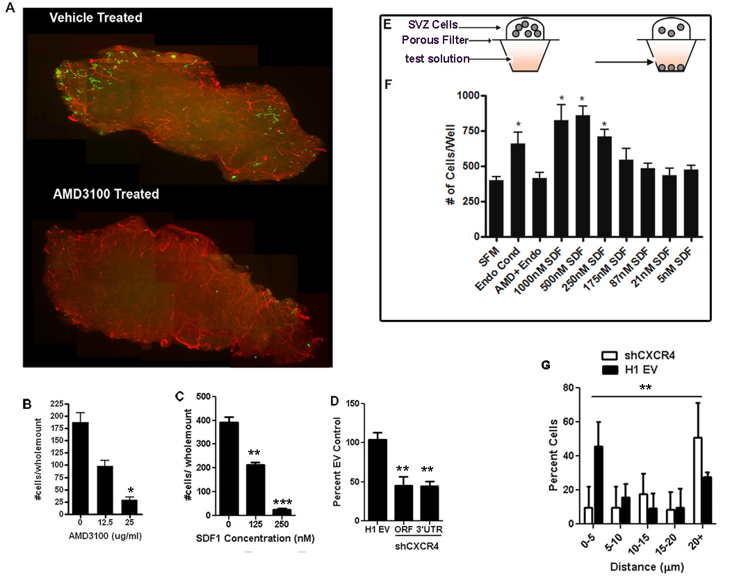
Figure 5.
SDF1/CXCR4 signaling is important for NPCs to locate the SVZ vasculature. GFP+ NPCs were overlaid onto SVZ wholemounts, incubated then fixed. The vasculature was stained for laminin (red). The number of cells entering the wholemount was counted (A,B) AMD3100 treated cells show significantly reduced ability to integrate into the wholemount, and this was dose-responsive. (C) SDF1 added to the media overlying the wholemount disrupts the gradient and inhibits cell integration. (D) Quantification of the number of transplanted SVZ cells entering the wholemount following lentiviral knockdown of CXCR4 receptor. (E) Schematic of the chemotaxis chamber assay used to measure attraction of NPCs to various media. (F) The number of cells that migrated towards control serum free media (SFM), endothelial conditioned media (Endo Cond), endothelial conditioned media with 25ug/ml AMD3100 (Endo + AMD3100) or various concentration of the chemokine SDF1 in the media(*p <0.05; **p <0.001; ***p< 0.0001). (G) Quantification of the distance of transplanted cells transduced with shCXCR4 or H1 empty vector to nearest blood vessel surface **p<0.001 on the distribution of shCXCR4 cells versus EV cells.

