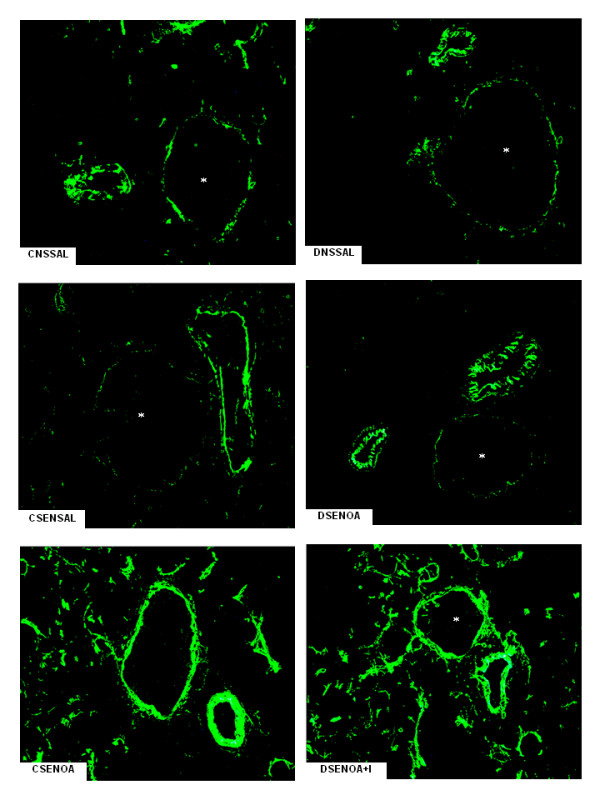
Figure 3.

E-selectin lung tissue microphotographs. The immune staining for E-selectin on lung microvessels of ovalbumin (OA) sensitized non-diabetic and diabetic rats 6 hours after OA (experimental) or saline (control) instillation. Insulin (NPH, 4 IU/rat s.c.) was given to diabetic rats 2 hours before OA challenge. Values are means ± SEM for 8 samples/rat, 3 animals/group. Analyses were performed by using the software image-pro Plus, version 4.1, Media Cybernetics. The microphotographs of lung tissue were obtained from control non-diabetic rats non-sensitized and instilled with saline (CNSSAL) or sensitized and instilled with saline (CSENSAL) or sensitized and instilled with OA (CSENOA), diabetic rats non-sensitized instilled with saline (DNSSAL) or sensitized and instilled with OA (DSENOA), and insulin treated diabetic rats sensitized instilled with OA (DSENOA+I). *Indicates the vessel lumen. Lung sections (8 μm) were stained for the detection of E-selectin (original magnification × 1500). *P < 0.001 comparing OA challenged with the control in the corresponding group (diabetic or non-diabetic). †P < 0.0001 comparing diabetic rats treated vs non-treated with insulin. ‡P < 0.001 comparing OA challenged with the diabetic or non-diabetic group.
