Fig. 4.
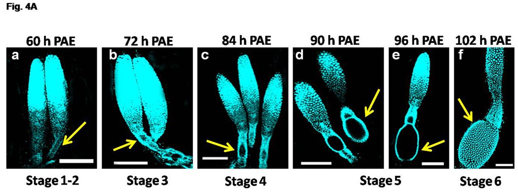
Fig. 4A. Progression of primary oocyte maturation during 60–102 h PAE. The ovaries were dissected at specific time-points and stained with nuclear stain, DAPI. The representative ovariole(s) at specific time-points are shown (panels a–f). The staging of the primary oocyte (yellow arrow) in the each ovariole was based on Ullmann (1973). The focus is on the staging of the primary oocyte. For distinct regions of the ovaries, see Fig. 4B. Note the increase in size of the primary oocyte at Stage 3 at 72 h PAE (panel b). The oocyte is spherical with follicular epithelial layer at Stage 4 at 84 h PAE (panel c). The oocyte becomes elongated surrounded by a monolayer of follicular cells at Stage 5 (panels d &e). Many follicular epithelial cells surrounding the mature oocyte are observed at Stage 6 (panel f). Scale Bar: 100 µm.
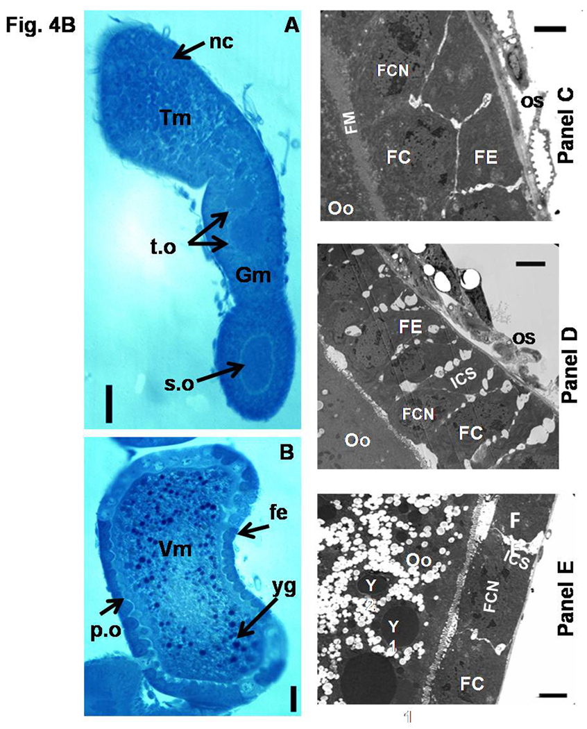
Fig. 4B. Light and Electron micrographs depicting the mature ovariole and the changes in the follicular epithelium of the primary oocytes (A–E). Light micrographs of oocytes/ovarioles stained with Methylene Blue. Semithin longitudinal sections of ovariole on day 5 PAE (A & B). Fe, follicular epithelium; nc, nurse cells; p.o, primary oocyte; s.o, secondary oocyte; t.o, tertiary oocyte; yg, yolk granules; Tm, tropharium; Gm, germarium; Vm, vitellarium.N=4 Scale Bar: A & B − 50 µm. Electron micrographs of a portion of the primary oocyte (C–E). Follicle epithelial cells organize into a columnar epithelium at Stage 5 (C). Note the intercellular spaces appearing between the follicle cells at Stage 6 (D). Yolk deposition was observed in the oocyte and the follicle cells flatten at Stage 7 (E). FC, Follicle cells; FE, Follicular epithelium; FCN, follicle cell nucleus; FM, Follicle membrane; ICS, Intercellular spaces; Oo, Primary oocyte; Os, ovarian sheath; Y, Yolk granules. Scale Bar: 2 µm.


