
Figure 4.
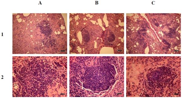
Histopathology of lung sections from BALB/c mice infected with wild type M. tuberculosis H37Rv (A), complemented mce1R mutant (B), and mce1R mutant (C). The mice were infected via the inhalation route and medicated for 4 weeks as described in Fig. 2. Lung sections from four mice per each group were harvested at 17 weeks post-medication (25 weeks post-infection) and stained with H&E. Lung tissues in all groups show a granulomatous interstitial pneumonia. Magnifications, × 100 (row 1) and × 400 (row 2); scale bars, 80 μm (row 1) and 20 μm (row 2).