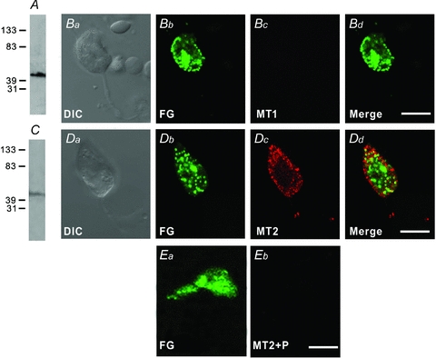
Figure 1. Expression of melatonin MT2 receptors in rat retinal ganglion cells (RGCs).

Double immunofluorescence labelling was performed using the antibody against MT1 or MT2 receptors on acutely dissociated RGCs labelled retrogradely by injecting fluorogold (FG) into the superior colliculus bilaterally. A, Western blot of whole rat retinal extract using the antibody against MT1, revealing a single band at the corresponding molecular weight of around 45 kDa. Ba–Bd, a RGC, double labelled by FG and MT1. Ba is the DIC image of the cell. The cell was labelled by FG (Bb), but not by MT1 (Bc). Bd is the merged image of Bb and Bc. C, Western blot of whole rat retinal extract using the antibody against MT2, also revealing a single band at around 45 kDa, corresponding to the molecular weight of the native MT2 receptor. Da–d, another RGC labelled by FG and MT2. Da is the DIC image of the cell. Dd is the merged image of Db, showing labelling for FG, and Dc, showing labelling for MT2. Note that the labelling for MT2 is primarily located on the membrane of the soma, and some punctate labelling is also observed in the dendrites. Ea, a RGC labelled by FG. Eb, no immunoflorescence labelling for MT2 receptors could be found when the MT2 antibody was pre-absorbed with the immunizing antigen. Scale bars, 10 μm.
