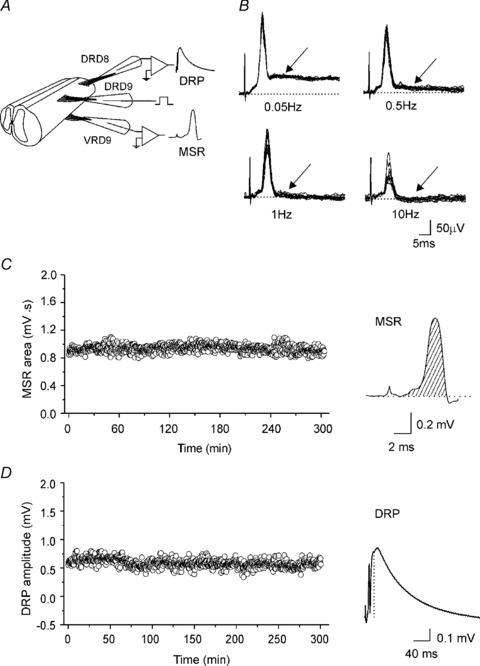
Figure 1. Simultaneous recordings of the monosynaptic reflex (MSR) and the dorsal root potential (DRP) in the turtle spinal cord.

A, scheme of two spinal cord segments in continuity with the dorsal and ventral roots attached to suction electrodes for stimulation and recording of the DRP and VRP. B, VRP evoked at four different frequency stimulations (2T) applied to the ipsilateral dorsal root. The earliest component of VRP is the MSR because it followed one to one frequency stimulation at 10 Hz without jittering and failure. The slower VRP component failed to follow one to one at 1 Hz of dorsal root stimulation. Striped area under the MSR curve (C) and the DRP amplitude (D) recorded simultaneously every 30 s plotted versus time. Traces, average of the MSR and DRP recordings of 10 sweeps.
