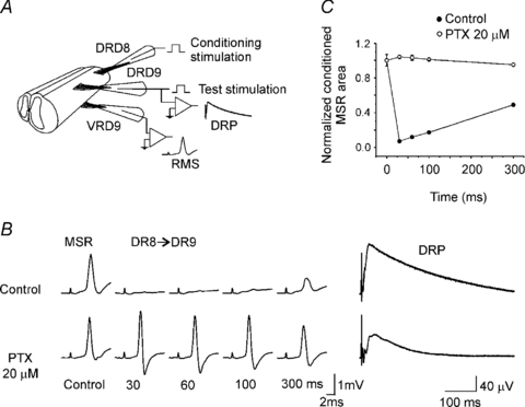
Figure 3. Long duration inhibition of the MSR.

A, scheme showing the protocol to induce long duration inhibition of the MSR. MSR recorded from D9 ventral root (VRD9) was evoked by stimulation of the ipsilateral dorsal root (DRD9). Conditioning stimulation was applied through D8 dorsal root (DRD8). B, MSR control and conditioned (DR8→DR9) recorded in control Ringer solution (top traces) and in presence of picrotoxin (20 μm; lower traces) at interstimulus intervals indicated bellow. To the right, DRP recorded from DRD9 in control Ringer solution and in the presence of picrotoxin. C, plot of normalized MSR area versus interstimulus interval in control Ringer solution (filled circles) and in presence of picrotoxin (open circles). Vertical bars indicate s.e.m.
