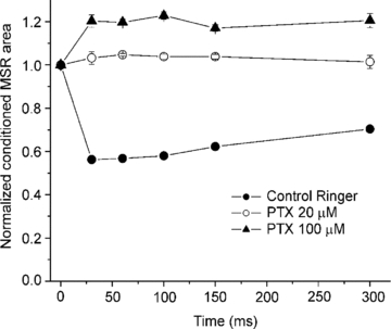
Figure 4. Faciliation of the conditioned MSR by higher concentrations of picrotoxin.

Plot shows the normalized area of MSR evoked by stimulation of the DRD9 conditioned by stimulation of an adjacent dorsal root (DRD8) at interstimulus intervals of 30, 60, 100, 150 and 300 ms in control Ringer solution (filled circles) and in the presence of picrotoxin at 20 (open circles) and 100 μm (filled triangles). Vertical bars indicate the s.e.m.
