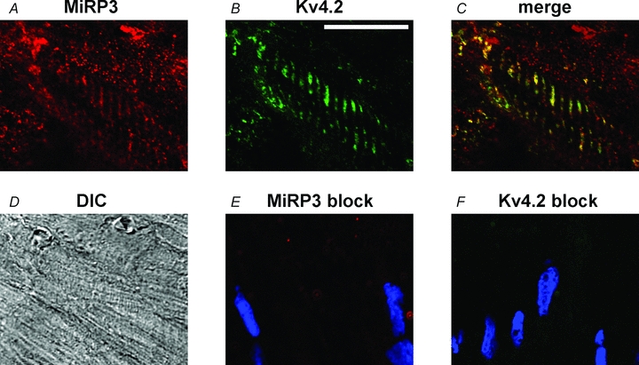
Figure 1. Spatial co-localization of MiRP3 and Kv4.2 within rat heart.

Confocal images of rat left ventricle probed with antibodies to MiRP3 (A) and Kv4 (B). The T tubules appear yellow in the merged image (C). A differential interference contrast image (DIC, D) is provided for reference. Images E and F show similarly obtained confocal images when antibodies to MiRP3 and Kv4.2, respectively, were preincubated with blocking peptide; the blue colour reflects DAPI staining of nuclei. The white scale bar in B represents 20 μm.
