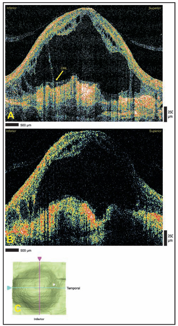
Figure 2.
Massive subretinal fluid due to exudative age-related macular degeneration. (A) Cirrus HD-OCT (Carl Zeiss Meditec, Dublin, CA) and (B) StratusOCT (Carl Zeiss Meditec) images demonstrating a large subretinal fluid accumulation overlying disciform scarring. The Cirrus HD-OCT image shows a portion of the outer plexiform layer (OPL) remaining adjacent to the scarred area. Note the finer detail of the cystoid structures displayed in the Cirrus HD-OCT image. (C) Interactive internal limiting membrane segmentation data map demonstrating the two-dimensional area of internal limiting membrane elevation. ONL = outer nuclear layer.

