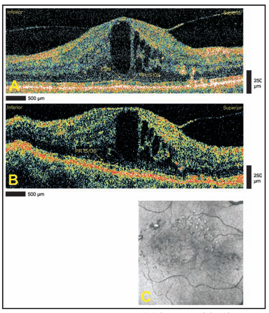
Figure 3.
Diabetic macular edema. (A) Cirrus HD-OCT (Carl Zeiss Meditec, Dublin, CA) and (B) StratusOCT (Carl Zeiss Meditec) images demonstrating cystoid edema. The Cirrus HD-OCT image demonstrates the continuity of the external limiting membrane (ELM) and the photoreceptor inner segment/outer segment (PR IS/OS) through the fovea. The ELM is not clearly seen in the StratusOCT image. (C) Advanced visualization C-scan image using a retinal pigment epithelium-aligned contour over a 53-µm thickness centered above the retinal pigment epithelium. Hard exudates are seen superiorly.

