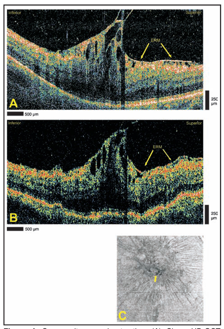
Figure 4.
Severe vitreomacular traction. (A) Cirrus HD-OCT (Carl Zeiss Meditec, Dublin, CA) and (B) StratusOCT (Carl Zeiss Meditec) images demonstrating vitreomacular traction and foveal cysts. An epiretinal membrane (ERM) is identified in both images. (C) Advanced visualization C-scan image using an internal limiting membrane-aligned contour over a 39-µm thickness centered at the internal limiting membrane. The arrow designates the location of foveal cysts.

