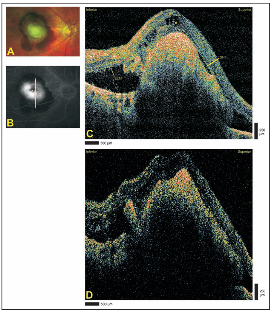
Figure 5.
Extensive choroidal neovascularization due to exudative age-related macular degeneration. (A) Optomap fa fundus photograph demonstrating subretinal hemorrhage. (B) Late-phase optomap fa fluorescein angiography demonstrating central hyperfluorescence surrounded by blockage by hemorrhage. Arrow depicts direction of optical coherence tomography scans. (C) Cirrus HD-OCT (Carl Zeiss Meditec, Dublin, CA) and (D) StratusOCT (Carl Zeiss Meditec) images demonstrating extensive choroidal neovascularization and subretinal hemorrhage. Superiorly, a small pocket of subretinal fluid (SRF) is identified in the Cirrus HD-OCT image, and the external limiting membrane (ELM) is seen in the inferior portion of the image. The asterisk designates cystoid edema.

