
Fig. 1.
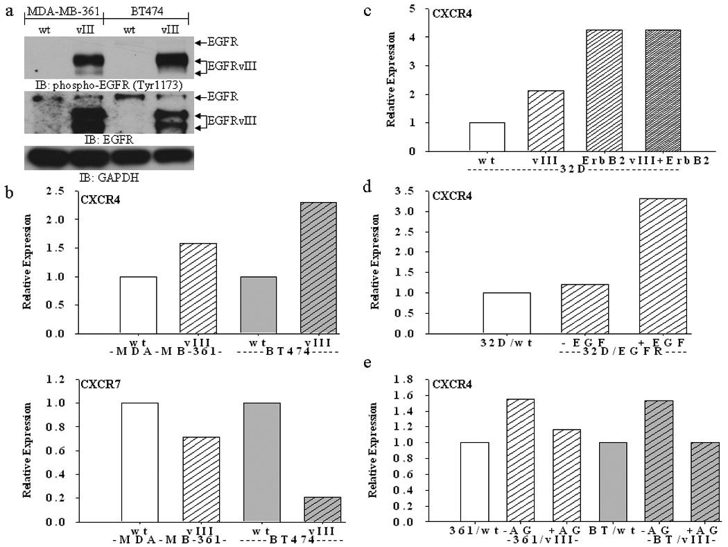
(a) Immunoblot analysis showing EGFRvIII is constitutively active in MDA-MB-361 and BT474 cells (parental cells are labeled “wt” and EGFRvIII-expressing cells are labeled “vIII”). (b) FACS analysis shows increased expression of CXCR4, but not CXCR7, in MDA-MB-361 and BT474 breast cancer cells stably expressing EGFRvIII. (c) FACS analysis shows increased expression of CXCR4 in 32D cells stably expressing EGFRvIII, ErbB2, and both EGFRvIII and ErbB2. (d) FACS analysis shows increased expression of CXCR4 in 32D cells stably expressing full-length EGFR (“32D/EGFR”) upon EGF (50 ng/mL) stimulation for 24 hours. (e) FACS analysis shows a reduction of CXCR4 in EGFRvIII-expressing MDA-MB-361 (“361”) and BT474 (“BT”) cells upon treatment with the TKI AG1478 (“AG”) (10 µM) for 24 hours. Bar graphs represent the relative expression based on the mean geometric fluorescence of cells in each group/control cells. Actual FACS plots are available in Supplemental Figure 1.