
Fig. 3.
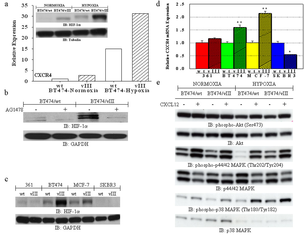
(a) FACS analysis shows increased expression of CXCR4 in BT474/vIII cells under both normoxic and hypoxic (24 hours) conditions in comparison to BT474/wt cells. Bar graphs represent the relative expression based on the mean geometric fluorescence of cells in each group/control cells. Actual FACS plots are available in Supplemental Figure 4. Inset, immunoblot analysis of lysates from BT474/wt and BT474/vIII cells under normoxic and hypoxic (24 hours) conditions showing increased expression of HIF-1α in BT474/vIII cells under both normoxic and hypoxic conditions. (b) Immunoblot analysis of lysates from BT474wt and BT474/vIII cells treated with the TKI AG1478 (10 µM) for 24 hours showing that inhibition of EGFRvIII activity can abolish increased HIF-1α expression in BT474/vIII cells under normoxic conditions. (c) Immunoblot analysis of lysates from MDA-MB-361 (“361”), BT474, MCF-7, and SKBR3 cells with and without EGFRvIII expression showing that HIF-1α is up-regulated in multiple breast cancer cells expressing EGFRvIII under normoxic conditions. (d) Quantitative real-time PCR analysis revealed significantly increased CXCR4 transcript levels only in BT474/vIII and MCF-7/vIII cells in comparison to parental cells (** = p<0.001; * = p<0.05, ANOVA). (e) Immunoblot analysis of lysates from BT474/wt and BT474/vIII cells showing that hypoxic conditions enhanced CXCL12-induced phosphorylation of Akt, p44/42 MAPK, and p38 MAPK. BT474/vIII cells also have higher basal levels of p44/42 MAPK and p38 MAPK activation under hypoxic conditions.