
Figure 1. The intracellular domain of HER4, s80HER4, inhibits growth via G2/M delay in HaLa-rtTA cells A.
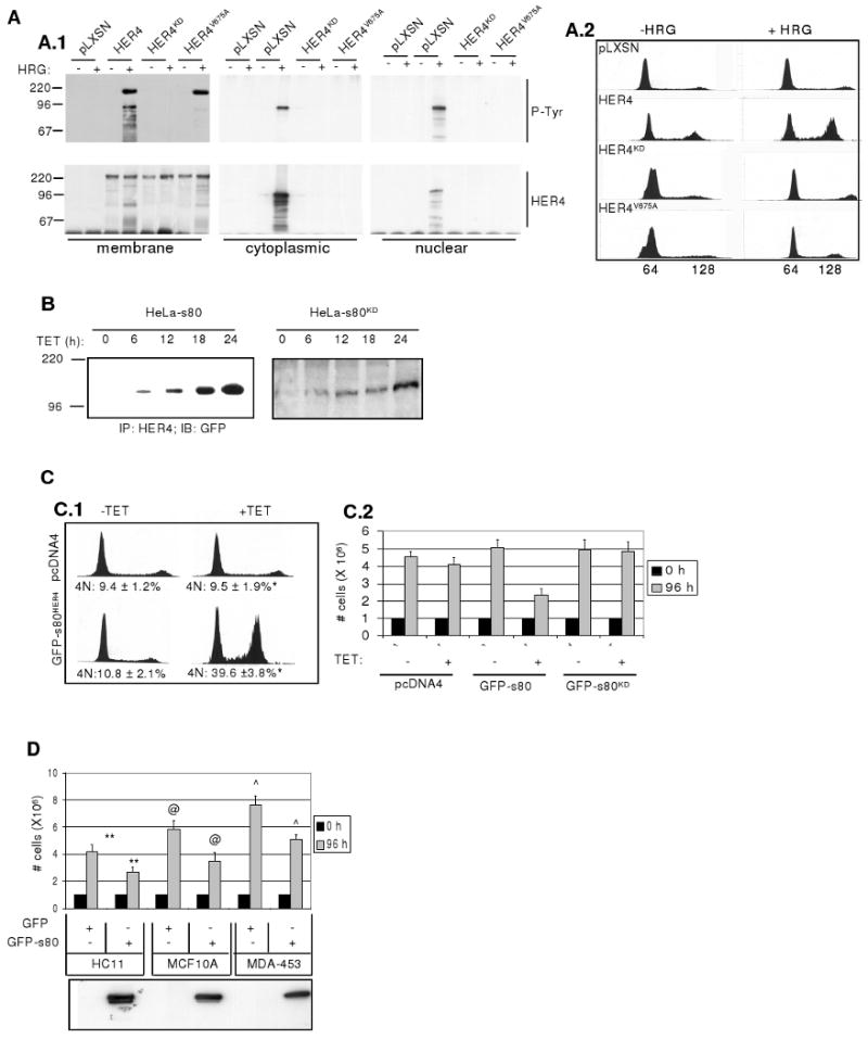
Mutation of the HER4 γ-secretase cleavage site inhibits formation of s80HER4 and interferes with HER4-mediated growth inhibition: A.1 Western analysis to detect phospho-tyrosine residues or total HER4 expression in HER4 immunoprecipitates (IPs) from membrane, cytoplasmic, or nuclear extracts from HeLa-rtTA cells expressing pLXSN, pLXSN-HER4, pLXSN-HER4KD, or pLXSN-HER4VA. Serum-starved cells were treated ± HRG (2h) before separation into cellular compartments using biochemical methods. Molecular weights are indicated at left. A.2: Cells grown in SF media ± HRG (48h) were stained with propidium iodide (PI) and analyzed by flow cytometry. Representative histograms are shown. B. Western analysis to detect GFP in HER4 IPs from HeLa-s80 and HeLa-s80KD cells cultured +TET for 0-24 h. C. Growth inhibition and G2/M delay in cells expressing GFP-s80HER4, but not in cells expressing GFP-s80KD. C.1: Representative histograms of HeLa-pcDNA4 and HeLa-s80 cells grown in SF media ±TET (48h), stained with PI and analyzed by flow cytometry. *P < 0.002, Student's unpaired T-test. C.2 Equal numbers of HeLa-pcDNA4, HeLa-s80, and HeLa-s80KD cells were plated, allowed to attach (24h), then treated for 0 or 96h ±TET in serum-free (SF) media. D. Equal numbers of HC11, MCF-10A, or MDA-453 cells expressing GFP-tagged s80 or GFP, were plated, allowed to attach (24h), then cultured for 0 or 96h in SF media. Each sample was counted in duplicate; each experiment was performed in triplicate. Values represent the average number of cells ± S.D. **P < 0.01; @P < 0.02; ˆP < 0.01; each calculated using Student's unpaired T-test Western analysis to detect s80HER4 in cell lysates is shown in lower panel.