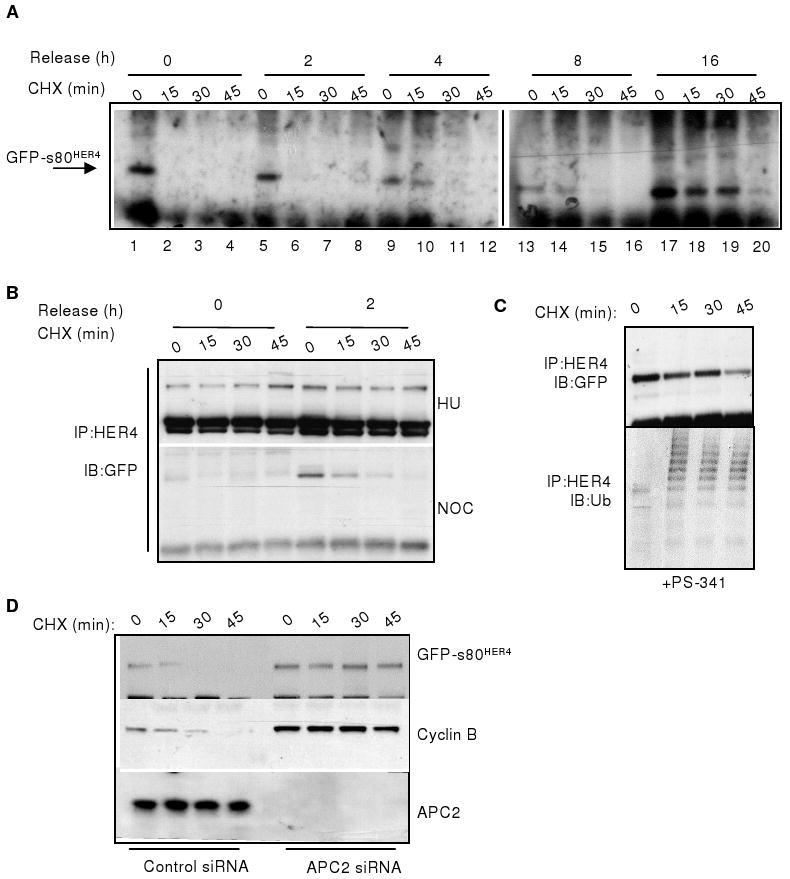
Figure 3. Degradation of s80HER4 during mitosis directed by APC/C. A.

HeLa-s80 cells were cultured +NOC/+TET in 10% serum for 16 h. Cells were released into 10% serum +TET at time (T) =0, and treated +CHX at T=0, 2, 4, 8, and 16h after NOC release. Expression of GFP-s80HER4 was analyzed at 15 min intervals beginning at the time of CHX treatment, by HER4 IP followed by GFP immunoblot (IB). B. HeLa-s80 cells were synchronized +HU/+TET (upper panel) or +NOC/+TET (lower panel) in 10% serum for 16 h, then released into 10% serum +TET at T=0. Cells were treated +CHX at T=0 and 2h. Expression of GFP-s80HER4 was analyzed as described above. C. Cells were cultured +NOC/+TET in 10% serum for 16 h, with PS-341 added for the final 4 h. Cells were released into 10% serum +TET/+PS-341 at T=0. CHX was added at T=0. Expression of GFP-s80HER4 was analyzed as described above; blots were stripped then probed for ubiquitin (lower panel). D. HeLa-s80HER4 cells transfected with 10 nmol siRNA sequences targeting luciferase or APC2 were treated +NOC/+TET +10% serum for 16 h. Cells were released into 10% serum +TET/+CHX. Expression of GFP-s80HER4 and cyclin B were analyzed at 15 min intervals beginning at the time of CHX treatment, by IB of anti-HER4 or anti-cyclin B IP's using GFP or cyclin B antibodies, respectively. APC2 was analyzed by IB.
