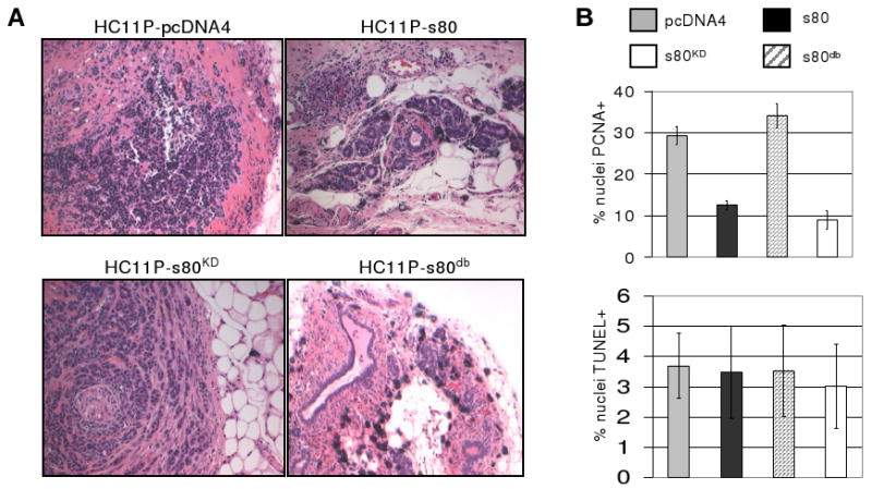
Figure 6. Decreased tumor formation in PyVmT-transformed HC11 cells expressing s80db. A.

HC11P cells expressing s80HER4, s80KD, s80db, or the empty pcDNA4 vector were injected into the inguinal mammary fatpads of female BALB/c mice. Fifteen weeks post-implantation of cells, mice were euthanized and the mammary glands analyzed histologically; representative images are shown. B. Five tumor samples per group were analyzed by immunohistochemistry. PCNA positive (upper panel) or TUNEL positive nuclei (lower panel) were scored in 3 randomly chosen 400× fields per tumor section, for a total of 15 fields counted. HC11P-pcDNA4 and HC11P-s80KD had equivalent percentages of PCNA-positive nuclei (29.4±2.2 and 34.1±2.9, respectively). HC11P-s80 had a statistically significantly lower percentage of PCNA-positive cells (12.5±1.1, p<0.0001) as compared to vector alone. While HC11P-s80db percentage of PCNA-positive cells was the lowest of all groups (9.0±2.2), and was also significantly decreased when compared to HC11P-s80 (p<0.037).
