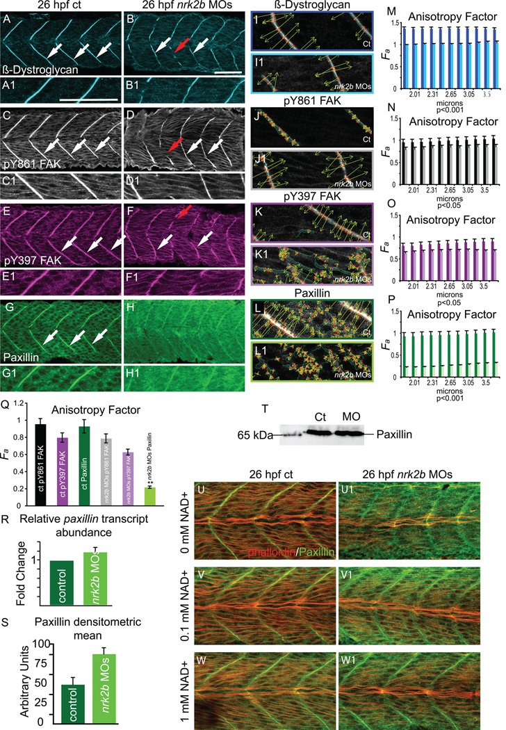
Fig. 6. Nrk2b is required for normal subcellular localization of Paxillin, but not β-Dystroglycan or FAK.
(A-H) Side mount, anterior left, dorsal top, 26 hpf. Panels A, C, E, and G are controls. Panels B, D, F, and H are nrk2b morphants. Panels numbered 1 are magnifications of the corresponding lettered panels. (A-B1) β-Dystroglycan antibody staining. (C-D1) pY861 FAK antibody staining. (E-F1) pY397 FAK antibody staining. (G-H1) Paxillin antibody staining. White arrows indicate normal localization of proteins to MTJs. Red arrows in nrk2b morphants (B, D, F) indicate discontinuous MTJs crossed by muscle fibers. Note that in nrk2b morphants (H, H1), Paxillin does not robustly concentrate to MTJs. (I-L) Side mount, anterior left, dorsal top, 26 hpf controls (lettered panels) and nrk2b morphants (numbered panels). Application of 2D WTMM method showing maxima chains (dark green), nodes (red), and vectors (light green) for CMAC component antibody staining. (M-Q) Graphs of anisotropy or the amount of order in a structure over a range of applicable size scales. (R) qRT-PCR, (S) average densitometric mean of Paxillin antibody-stained images and (T) Western analysis suggest that Paxillin levels are not decreased in nrk2b morphants. (U-W) Side mount, anterior left, dorsal top, 26 hpf embryos, phalloidin staining in red, Paxillin antibody staining in green. Lettered panels are controls and numbered panels are nrk2b morphants. (panels U) No exogenous NAD+. (panels V) 0.1 mM exogenous NAD+. (panels W) 1 mM exogenous NAD+. Note that exogenous NAD+ treatment rescues medial Paxillin concentration at the MTJ in nrk2b morphants in a dose dependent manner. Scale bars are 50 µm.

