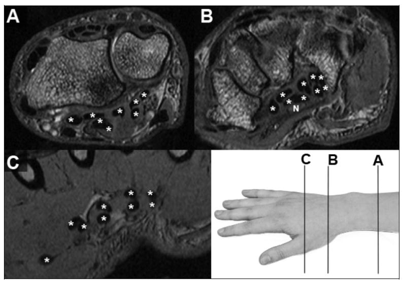
Figure 1.

MR images, with flexor tendons identified (*) within (B) and on either side of (A&C) the carpal tunnel. Only in the distal image from the hand (C), where the tendons align in pairs going to each finger (one to the thumb) and in two layers (deep and superficial), is unambiguous tendon identification possible. The median nerve (N, in panel B) has stippled image texture of intermediate signal intensity.
