
Figure 4.
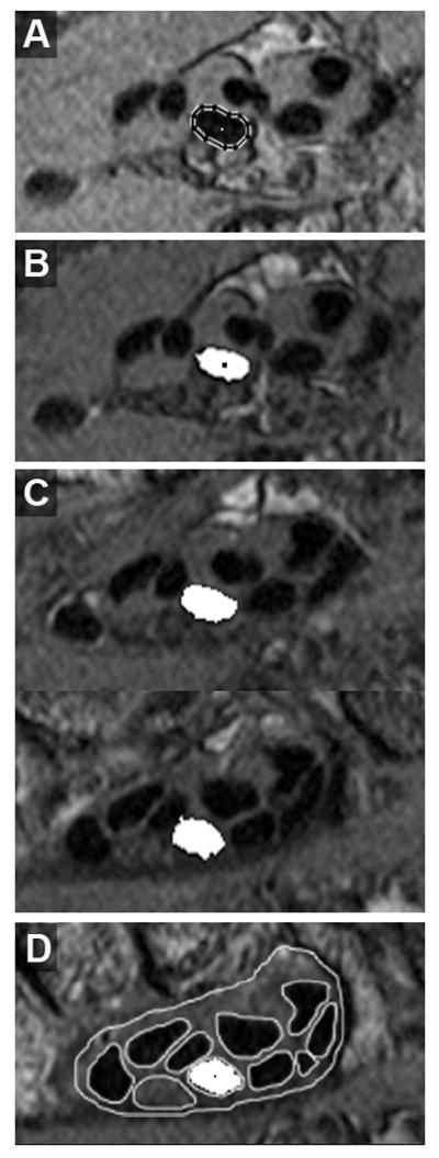
Progressive automatic tracking of the 3rd digit superficial tendon, beginning from the starting image. The centroid of the outlined tendon boundary (A) is used to establish the seed region (B) from which the next tendon region is grown. Additional cross-sections of this tendon at six-section increments, moving proximally toward the distal end of the carpal tunnel are shown in C. Tracking continues until the image of the distal carpal tunnel is reached, which coincides with the distal-most segmentations (D).