
Fig. 3.
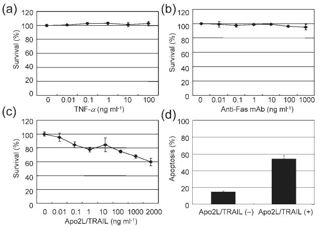
Apo2L/TRAIL-induced cytotoxicity and apoptosis. (a)–(c) HepG2 cells were incubated with various concentrations of TNF-α (a), agonistic anti-Fas mAb (b) or Apo2L/TRAIL (c) for 72 h. The WST-8 assay was used to measure cell viability. Results are shown as percentage survival compared with the control. (d) The proportion of Apo2L/TRAIL (500 ng ml−1)-treated HepG2 cells in an apoptotic state, as determined by Apo2.7 staining. Values represent the mean±SD of triplicate measurements.