
Figure 5.
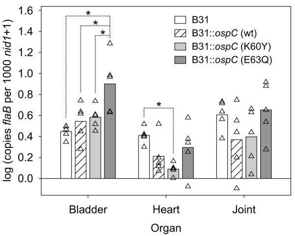
Quantitative PCR analysis of spirochete burdens in bladder, heart, and joint of infected mice. Mice were infected by needle inoculation and after 4 weeks organs were harvested. DNA was isolated from each organ and spirochete burden determined by amplification of the B. burgdorferi flaB gene (normalized to copies of the mouse nid1 gene). All analyses were repeated three times (in triplicate each time). Results from individual mice are shown as triangles, with the mean denoted by the bar. Statistical significance (p<0.05) between specific data sets (indicated by the lines above the bars) is denoted by *.