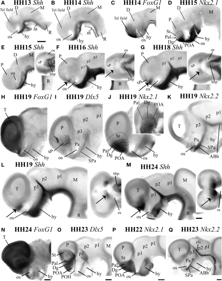
Figure 1.
The Shh-positive domain appears in the subpallium at HH16, being strictly separate from the Shh-positive basal plate domain. Whole mount in situ hybridization for Shh in the early developing embryo in lateral views at HH13 (A), HH14 (B), HH15 (E), HH16 (F), HH18 (G), HH19 (L), HH24 (M) are compared with whole mount in situ hybridization for the telencephalic marker FOXG1 at HH14 (C), HH19 (H), and HH24 (N), the subpallial marker Dlx5 at HH19 (I) and HH23 (O) and the pallidal marker Nkx2.1 at HH15 (D), HH19 (J), and HH22 (P). The alar-basal boundary was visualized with in situ hybridization for Nkx2.2 at HH19 (K) and HH23 (Q). The insets at the top right corner of (E), (F), (G), (J), (L), and (M) are ventral views at the same magnification as the respective lateral views. The arrows in (F), (G), (L), (M) indicate in lateral or ventral views the Shh-positive domain detected in the subpallial preoptic alar region. Scale bar in (A) = 0.2 mm applies to (A)–(L), whereas the bar = 0.3mm in (M)–(Q).

