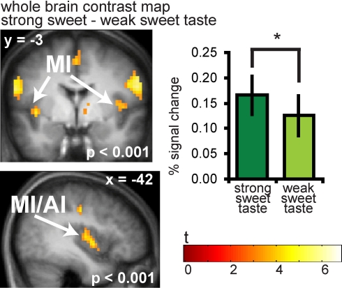
Figure 3.
Neural response in the insula to sweet taste. Coronal and sagittal sections of the insula showing response to strong sweet taste – weak sweet taste. In the bargraphs we plotted the average percent signal change for the two sweet taste conditions over subjects (±s.e.m.): (Figure 6A and 6B) strong sweet taste (dark green) and weak sweet taste (light green), averaged over subjects. The response was taken from the voxel that responded maximally, as identified in the SPM analysis. Thus, these estimates are non-independent, and are provided to illustrate the relative difference in neural response.

