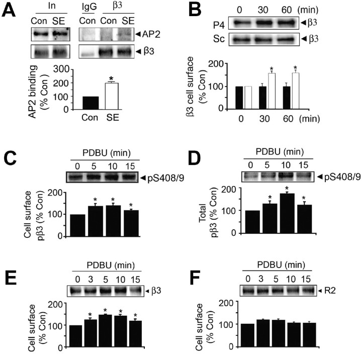
Figure 5.
Blocking endocytosis and the activation of PKC increases GABAAR cell surface expression levels in SE. A, Increased association of GABAARs with AP2 in SE. Detergent-solubilized extracts were immunoprecipitated with IgG or anti-β3 antibodies, and precipitated material was immunoblotted with antibodies against the GABAAR β3 subunit or the α subunit of AP2. In represents 20% of the input used for each immunoprecipitation. After controlling for recovery of the β3 subunit, the level of AP2 binding was determined for control (filled bar) and SE (open bar), and data were normalized to levels evident in control (100%). B, Blocking endocytosis increases GABAAR cell surface stability in SE. Hippocampal slices from SE animals were incubated in the presence of 90 μm P4 or control Sc peptide. Top, Slices were then subjected to biotinylation with NHS-biotin, and cell surface fractions were immunoblotted with anti-β3 antibody. Bottom, The levels of cell surface β3 in slices exposed to either Sc (filled bars) or P4 (open bars) were then normalized to those evident at 0 time (100%). C, D, PKC activators increase GABAA receptor phosphorylation and cell surface expression levels in SE. Slices were incubated with 100 nm PDBU and labeled with NHS-biotin. Cell surface (C) and total fractions (D) were then immunoblotted with pS408/9 antibody. The level of phosphorylation of S408/9 was normalized to that evident at 0 time (100%). E, F, PKC activators increase GABAA receptor cell surface stability in SE. Cell surface fractions from SE slices were blotted with anti-β3 (E) and antibodies against the GABABR2 subunit (F), and levels of each protein were normalized to those seen at 0 time (100%). In all panels, asterisks indicate significant difference from control (p < 0.01; Student's t test; n = 5–7).

