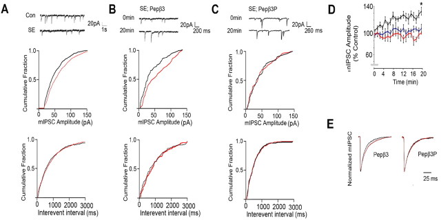
Figure 6.
Blockade of endocytosis increases the amplitude of mIPSCs and slows their decay in CA1 neurons undergoing SE. A, SE reduces mIPSC amplitude. Top, Representative sweeps. Middle, bottom, Cumulative probability analysis for mIPSC amplitude (middle) and frequency (bottom; interevent interval) recorded from CA1 neurons in control (black) and SE (red) slices. B, C, Pepβ3 selectively enhances mIPSC amplitude in SE neurons. Top, Representative sweeps at 0 and 20 min for CA1 neurons exposed to 100 μg/ml Pepβ3 (KTHLRRRSSQLK; B) or Pepβ3P (KTHLRRRSPSPQLK; C) via intracellular dialysis. Cumulative probability for mIPSC amplitude and frequency are also shown at 0 time (red) and 20 min (black), respectively. D, Time-dependent enhancement of mIPSCs by Pepβ3. mIPSC peak amplitude from CA1 neurons dialyzed with control (blue), Pepβ3 (black), and Pepβ3P (red) over a 20 min time course was normalized to that seen at 0 time (100%). E, Pepβ3 modulates mIPSC decay. Scaled mIPSCs are shown for CA1 neurons from SE slices dialyzed with Pepβ3 or Pepβ3P at 0 (black) and 20 min (red), respectively. In all panels, asterisks indicate significant difference from control (p < 0.01; Student's t test; n = 8–12).

