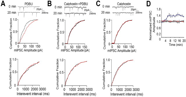
Figure 7.
PKC-dependent activity modulates mIPSCs in SE. A–C, PKC activity enhances mIPSC amplitude in SE. Representative sweeps from CA1 neurons exposed to PKC activators/inhibitors and cumulative amplitude distributions for events at 0 time (red) and 20 min (black) are shown. D, PDBU enhances mIPSC amplitude in SE. Peak amplitudes were measured over a 20 min recording period for SE CA1 neurons treated with PDBU (100 nm; blue), PDBU/calphostin (100 nm and 10 μm, respectively; red), and calphostin alone (10 μm; black), and data were normalized to those seen at 0 time (100%).

