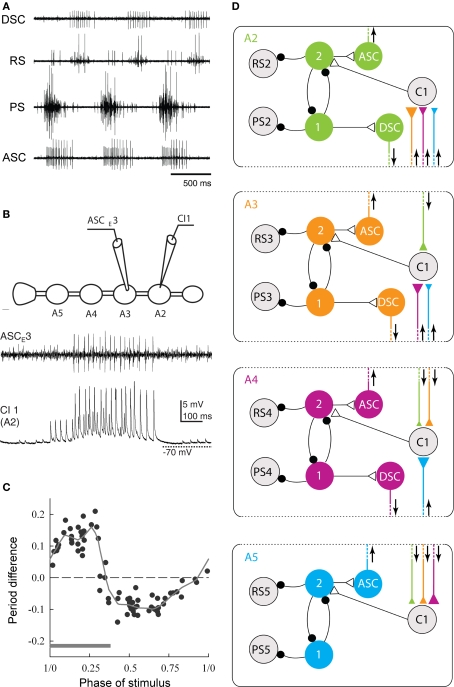
Figure 3.
The neural circuit that coordinates swimmeret movements. (A) Simultaneous recordings from PS and RS branches of a nerve innervating one swimmeret and from axons of coordinating neurons that project from the local circuit that controls firing of the swimmeret's motor neurons. Bursts of spikes occur in the anterior-projecting coordinating axon (ASC) simultaneously with each PS burst. These bursts alternate with bursts in the posterior-projecting axon (DSC) that occur simultaneously with RS bursts. Each burst in ASC or DSC encodes when the corresponding burst of spikes in motor neurons began, how long it lasts, and how strong it is (Mulloney et al., 2006). (B) Simultaneous recordings of a burst of spikes in an ASC coordinating axon and the EPSPs in a ComInt 1 commissural neuron (C1) onto which the ASC axon synapses. The cartoon shows the positions of the two recording electrodes on ganglia of the abdominal nerve cord preparation. A2, A3, A4, A5: the second through fifth abdominal ganglia, each of which innervates a pair of swimmerets. (C) A phase-response curve that shows how the phase at which a pulse of depolarization in a ComInt 1 neuron affects the period of the local circuit's output. Here, phase is defined as the point in the cycle of PS–RS alternation at which the stimulus began. Stimuli early in a cycle, during the PS burst (gray bar), prolong the period of the cycle, but later stimuli shorten the period. The period difference is the difference between the measured period and the expected period if the stimulus had no effect. The dotted horizontal line marks zero change. The solid line is a “locally weighted estimate” fit to the data (Smarandache et al., 2009). (D) A diagram of the circuit that coordinates local pattern-generating circuits in ganglia A2, A3, A4, and A5. In each ganglion, local interneurons (1, 2) determine when the PS and RS motor neurons fire. In each ganglion, an ASCE and a DSC neuron encode information about the PS and RS activity and conduct it to other ganglia. This information converges onto ComInt 1 neurons (C1) that synapse onto one of the local pattern-generating neurons. Colors mark the ganglion in which each coordinating axon originates. Triangles symbolizes excitatory synapses; larger triangles mark relatively stronger synapses. Small black circles symbolize inhibitory synapses. Arrows show the direction of impulse conduction in each coordinating axon.

