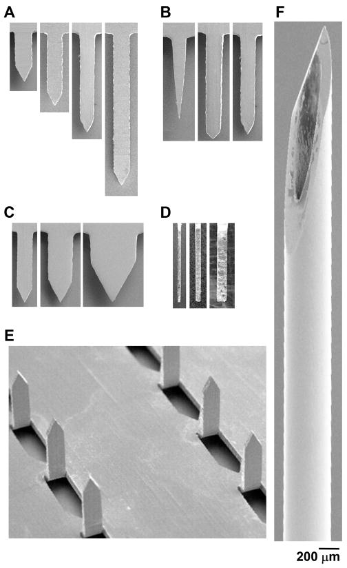
Figure 2.
Representative microneedles used for insertion. Scanning electron microscopy images of microneedles used in the length study (A), tip-angle study (B), width study (C), thickness study (side view) (D) and number of microneedles study (E). A 5 mm-long, 26-gage hypodermic needle was used as a positive control (F). All images are at the same magnification.

