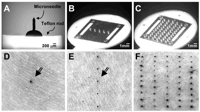
Figure 3.

Microneedle devices and stained skin penetration sites. Brightfield microscopy images of microneedles assembled into devices: a single microneedle affixed to a teflon rod holder (A), a five-microneedle array assembled as an adhesive patch (B) and a 50-microneedle array assembled as an adhesive patch (C). Brightfield microscopy images of the skin surface of human forearms after inserting microneedles and applying gentian violet to stain the sites of microneedle insertion, which demonstrates microneedle penetration into the skin, using: a single microneedle (D), an array of five microneedles (E) and an array of 50 microneedles (F). Arrows in (D) and (E) point to the stained insertion sites.
