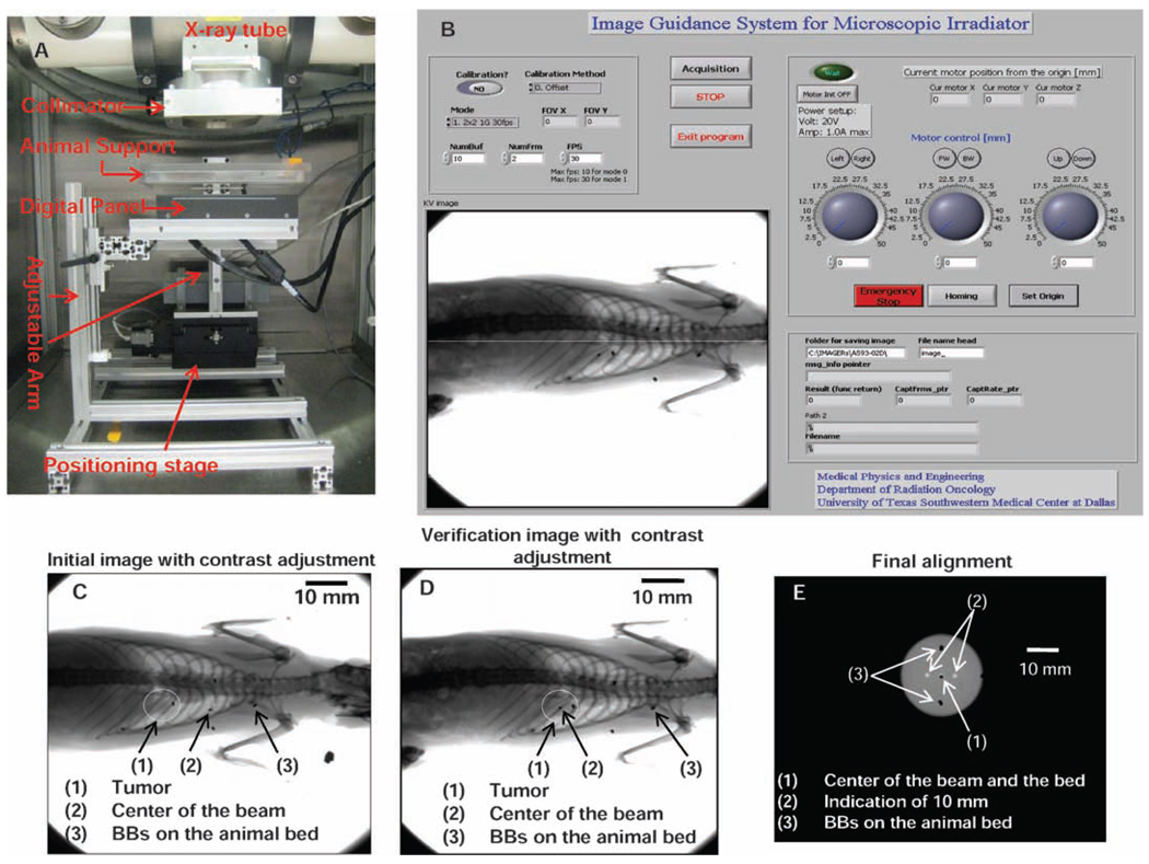
FIG. 1.
The image-guided small animal irradiation device (panel A) with major components indicated, positioned beneath a commercial X-ray source. Panel B: Software for image acquisition and positioning. Panel C: Initial localization image with beam center and tumor indicated. Panel D: Final localization verified by a second image. Panel E: Calibration and centering relative to the beam axis.

