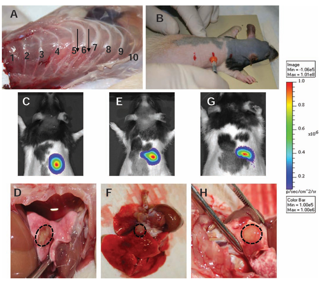
FIG. 2.
Development of rat orthotopic model for SBRT. Panel A: Dissected specimen showing the desired injection site. Panel B: Rats are anesthetized using rat cocktail and placed on a supporting frame head up inclined at 30° angle. The upper, middle and bottom marks indicate ribs 1, 6 and 12, respectively. Cells mixed with matrigel are implanted using a 28G1/2 needle. A needle guard is used to control the depth. Panels C, E and G: Bioluminescence imaging in three animals 3 weeks after implantation. Panels D, F and H: Corresponding high-resolution digital image of the solitary nodule in the rat lung.

