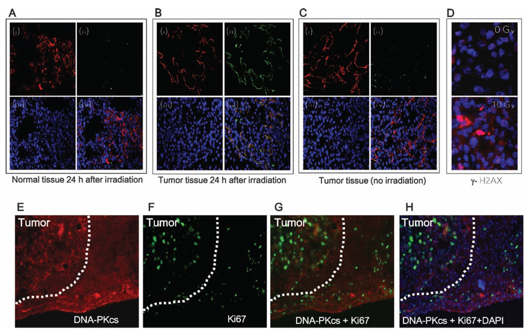
FIG. 4.
Target validation after Image-guided radiation. Panel A: Normal lung tissue harvested 24 h after irradiation; panel B: tumor tissue harvested 24 h after irradiation; panel C: Unirradiated tumor tissue. In all panels: (i) vascular endothelium detected by mouse anti-rat CD31 antibody followed by Cy3-goat anti-mouse antibody (red), (ii) bavituximab detected using biotinylated goat anti-human IgG followed by Cy2-streptavidin (green), (iii) DNA detected by DAPI (blue), (iv) CD31, bavituximab, and DNA merge image. Panel D: Tumor immunofluorescence staining; merged image of DAPI and phospho-γ-H2AX (0 Gy, upper panel) and irradiated tumors (10 Gy, lower panel). Panel E: DNA-PKcs; panel F: Ki67; panel G, DNA-PKcs and Ki67 merge image; panel H: DNA-PKcs, Ki67 and DAPI merge image.

