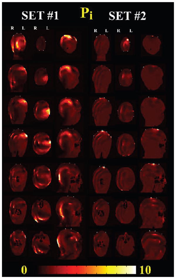
FIG. 4.

Changes in RF-field power with and without leads (Pi) for 915 MHz mobile phone exposure with the two sets of EEG electrodes/leads modeled. Coronal (from the back to the front of the head), axial (from the top of the head to the neck), and sagittal (from right to the left of the head) are shown. The phone was placed on the left of the head. Significant increases in Pi were observed for Set 1 near the EEG leads and in all the anatomical structures underneath the EEG electrode near the source (coronal and axial view) and on the back of the head near the leads (sagittal view). Changes for Set 2 were localized mainly underneath the electrode in Cz position, where the leads were bundled.
