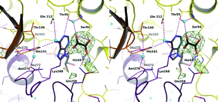
Figure 2.
Stereoview of the OMIT map of oxalate and interactions in the active site. The OMIT F o − F c difference density map is shown as green chicken wire and contoured at 3σ. The protein main chain is shown in ribbon format coloured according to subunit: yellow, subunit A; orange, subunit B; purple, subunit D. Side chains are shown as sticks coloured red for O atoms, blue for N atoms, orange for P atoms and according to subunit as before for C atoms. The oxalate and AMP C positions are shown in black. Water molecules are depicted as cyan spheres. Dashed lines represent potential hydrogen-bonding interactions.

