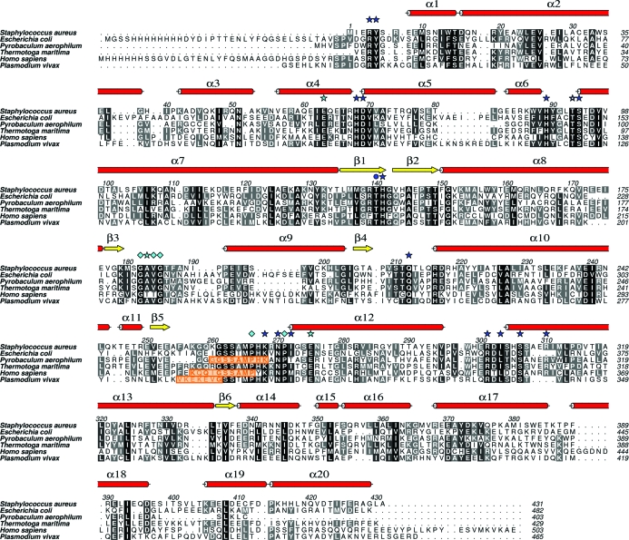
Figure 4.
Primary and secondary structure of SaPurB together with sequence alignment of selected PurB sequences. Dark blue stars indicate residues that interact with ligands in the active site; a dark blue circle identifies a threonine previously thought to be important for activity and discussed in the text. Light blue stars mark residues that help to position important side chains; light blue diamonds indicate structurally important residues. The orange boxes highlight disordered regions that correspond to the mobile active-site loop in SaPurB.

