Figure 3.

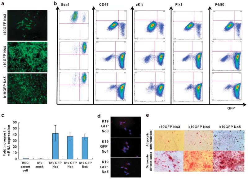
Establishment of GFP-labeled MSC clones, that express K19. (a) GFP expression in MSC clones established by transfection with a K19–EGFP gene construct. After selection with G418 treatment, 3 of 11 isolated clones expressed GFP and were designated as K19GFP no. 3, no. 4, and no. 5. (b) Expression of GFP and cell surface markers (Sca1, c-kit, CD45, Flk1, and F4/80) in GFP-positive clones was analyzed by flow cytometry. Cells were stained with PE-conjugated antibodies, and both antibody staining and endogenous GFP expression was detected. Quadrant markers were set according to the profile of control IgG staining in GFP-negative parent cells. Representative examples of three experiments are shown. (c) Fold increase in K19 mRNA expression level in mock transfectant, K19GFP no. 3, no. 4, and no. 5 is shown, as compared with that of parent cells by RT-PCR (n = 3). (d) K19 protein expression in GFP-positive clones was assessed by immunofluorescence staining with a Texas Red conjugated anti-K19 antibody. Nuclei were stained with DAPI. Original magnification, × 100. (e) Osteocyte and adipocyte differentiation 14 days after incubation with appropriate culture condition in K19GFP no. 3, no. 4, and no. 5 were detected by Alizarin Red and Oil red-O staining, respectively. Original magnification, × 40.
